
Tecpr2 Mutation Impairs Endolysosomal Activity in Neurons and Microglia
A recent research study published in Cell Death & Disease, successfully integrated whole-animal phenotypic data (behaviour, physiology, histopathology) with cellular and molecular analyses to investigate hereditary sensory and autonomic neuropathy subtype 9 (HSAN9), a fatal neurodevelopmental and neurodegenerative disorder.
Systemic phenotyping of Tecpr2 knock-in (ki/ki) mice, which mimic the nonsense mutation L1139Rfs and show loss of TECPR2 expression, revealed altered gait and neurodegenerative changes in the medulla oblongata of the brainstem.
Behavioural assays (open field, repulse inhibition and CatWalk) in Tecpr2 ki/ki mice, used to characterize aspects of emotionality, sensorimotor function and coordination, clearly demonstrated decreased anxiety-related behaviour, impaired sensorimotor gating and increased walking cadence. These mice displayed reduced temporal and spatial walking parameters as well as decreased forelimb footprint area (Figure 1).
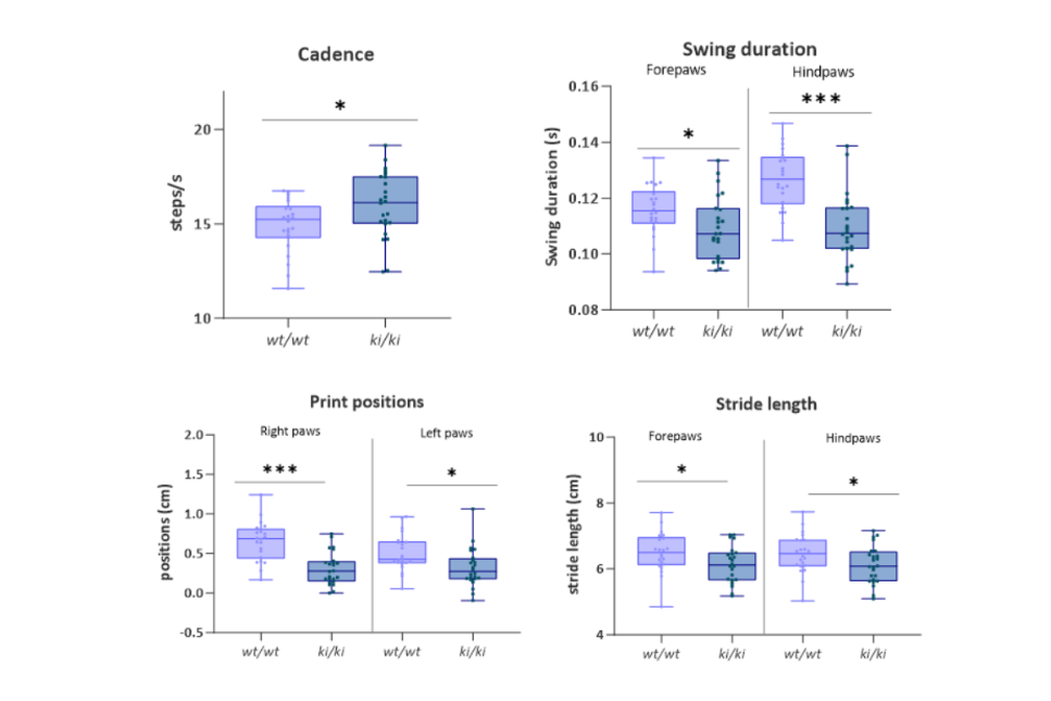
Figure 1: Temporal and spatial walking parameters
A detailed histopathological evaluation of the brain revealed morphological alterations exclusively in the medulla oblongata of Tecpr2 ki/ki mice (Figure 2). These changes included swollen hypereosinophilic axons (axonal ‘spheroids’), a reduction in neuronal cell bodies, extensive loss and fragmentation of neurofilaments and a decrease in oligodendrocytes. Furthermore, the medulla of mutant mice showed increased numbers of astrocytes and enlarged microglial cells with ramified processes, indicative of astrogliosis and microgliosis. In addition, IF for autophagosomal, endolysosomal and synaptic vesicle markers indicated impairment of autophagosomal-endolysosomal pathways.
Proteomics and electron microscopy of the affected medulla region revealed altered organelle and protein homeostasis, characterized by the accumulation of amorphous protein aggregates, glycogen granules, autophagosomes and synaptic vesicles as well as enlarged endoplasmic reticulum and swollen mitochondria whose proteomes were also altered.
The study further investigated TECPR2 knockout cells to better understand the impact of TECPR2 on the endolysosomal system in neurons and microglia.
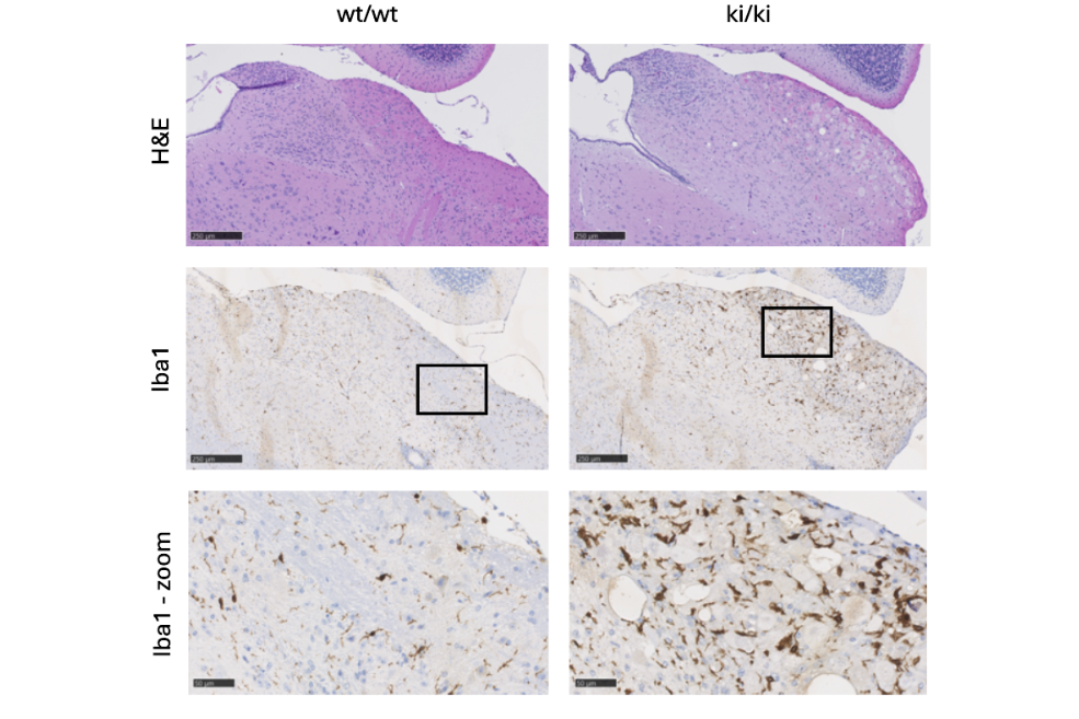
Figure 2: Histological evaluation of brain tissue
In summary, this study provides molecular and pathophysiological insights into the role of TECPR2 in the endolysosomal system. Behavioural and phenotypic analyses of Tecpr2 ki/ki mice show a gait ataxia phenotype similar to that observed in HSAN9 patients and axonal dystrophy consistent with findings in Tecpr2 knockout mice. While histological phenotyping of Tecpr2 ki/ki mice confirms that loss of TECPR2 causes neurodegeneration, it also illustrates that other brain cell types, in particular microglia, are affected for example in their ability to clear endocytic and phagocytic cargo which may in turn contribute to disease progression.
Original publication:
Bhattacharya, D., da Silva-Buttkus, P., Nalbach, K. et al. Neuropathy-associated Tecpr2 mutation knock-in mice reveal endolysosomal loss of function phenotypes in neurons and microglia. Cell Death Dis 16, 775 (2025). https://doi.org/10.1038/s41419-025-08168-w.